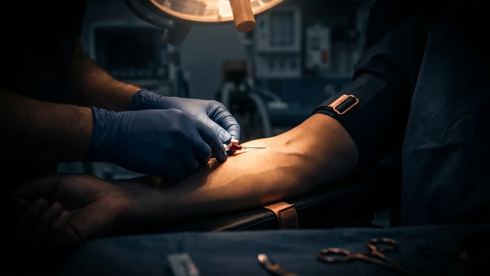
IV phlebitis signs and treatment start with recognizing vein inflammation early, grading it with a Visual Infusion Phlebitis (VIP) score, removing the catheter when indicated, and treating the site while you prevent the next case through better cannulation choices. The red streak along a vein is not a mystery. It is a clinical signal you can grade and act on mid-shift.
If you already know IV infiltration signs and treatment and IV extravasation management, this article fills the remaining major peripheral complication: inflammation of the vein itself. Different problem. Different grading tool. Same principle. Catch it early, respond cleanly, and fix the insertion habits that keep causing it.
What is IV phlebitis?
IV phlebitis is inflammation of the vein wall associated with a peripheral intravenous catheter, usually from mechanical irritation, chemical irritation, or bacterial contamination. Patients feel pain or tenderness along the vessel. You see erythema, warmth, swelling, and in later stages a palpable venous cord. When clot forms with the inflammation, clinicians often call it thrombophlebitis.
Phlebitis rates for peripheral IVs vary widely by definition and surveillance method, but many units treat sustained rates above about 5% as a quality problem once a validated tool like the VIP score is used consistently. Andrew Jackson developed the VIP score to standardize bedside recognition, and later validation work (including Gallant and Schultz, 2006) supported its use for deciding when a PIV should come out.
This is educational clinical reference content, not a substitute for your facility protocol or a medical order.
IV phlebitis signs you should not ignore
Early signs are easy to dismiss as "the patient is just sensitive." Do not.
Early signs
- Slight pain or tenderness near the insertion site
- Mild erythema at or around the catheter entry
- Patient reports that the site "burns" or "feels warm" during infusion
Progressive signs
- Pain that tracks along the path of the vein
- Red streak formation proximal to the insertion site
- Localized swelling and induration (firmness)
- Warmth along the vessel course
- Slowed infusion without a clear occlusion elsewhere
Advanced signs
- Palpable venous cord under the skin
- Extensive erythema and swelling
- Purulent drainage at the site
- Fever (pyrexia) with progressive local findings
Red streak recognition matters because nurses search for it when the arm already looks wrong. A linear erythema following the vein is classic mechanical or chemical irritation progressing. Treat the finding as a VIP reassessment trigger, not a wait-and-see decoration.
Mechanical, chemical, and bacterial phlebitis
Naming the type guides prevention. Treatment still starts with remove-and-support for most peripheral sites.
| Type | Typical trigger | What you notice | Prevention focus |
|---|---|---|---|
| Mechanical | Catheter too large for the vein, poor securement, joint-site placement, trauma during insertion | Pain with movement, early erythema, cord later | Smaller appropriate gauge, securement, avoid AC when possible |
| Chemical | Irritating drugs, high osmolarity, extremes of pH, rapid infusion of vesicants/irritants | Burning during infusion, streak along vein | Dilution, rate control, appropriate device for therapy |
| Bacterial | Breaks in aseptic technique, contaminated hub/site, prolonged dwell with poor care | Later onset, drainage, fever, systemic signs | Aseptic insertion and hub care, timely removal |
Bottom line: Mechanical and chemical phlebitis are often insertion and device-selection problems. Bacterial phlebitis is an infection-control problem. All three look similar early. Grade the site. Do not guess the cause from one glance and leave a VIP 2 catheter in place.
The VIP score for IV phlebitis grading
The Visual Infusion Phlebitis score is a 0 to 5 bedside scale. Score the site each assessment. Document the findings that justify the number, not just the number alone.
| VIP score | Clinical picture | Action |
|---|---|---|
| 0 | Site appears healthy | Observe cannula |
| 1 | One early sign: slight pain near site OR slight redness | Observe / consider resite if risk factors rise |
| 2 | Two of: pain, erythema, swelling | Resite cannula |
| 3 | Pain along path of cannula, erythema, and induration | Resite; consider treatment |
| 4 | Grade 3 findings plus palpable venous cord (advanced phlebitis / early thrombophlebitis) | Resite; initiate treatment; notify per protocol |
| 5 | Grade 4 findings plus pyrexia (advanced thrombophlebitis) | Initiate treatment; resite; notify provider; escalate |
A practical rule many units enforce: VIP 2 or higher means the catheter comes out. Waiting for a cord or fever is waiting too long. Follow your facility policy when it is stricter.
IV phlebitis signs and treatment at the bedside
When findings confirm vein inflammation, run a short protocol. Do not improvise under pressure.
- Stop the infusion and assess the VIP score. Confirm this is phlebitis, not infiltration (cool swelling of tissue) or extravasation (vesicant leak with tissue-injury risk).
- Remove the catheter when indicated. For VIP 2 and above, resite. Do not "finish the bag" through an inflamed vein.
- Elevate the limb. Reduces local congestion and patient discomfort.
- Apply a warm, moist compress. Typically 20 minutes, several times daily, unless your protocol specifies otherwise. Warmth supports comfort and local circulation for sterile inflammation.
- Support comfort and inflammation control per orders. Oral or topical anti-inflammatory options may be used when not contraindicated. This is a provider decision, not a default nurse add-on.
- Restart access thoughtfully. New site, preferably other extremity or proximal on a different vein pathway. Match gauge and vessel size. Avoid restarting distal to a damaged segment on the same vein.
- Document and report. Grade, findings, drug infusing, dwell time, actions taken, and patient response. Many facilities require incident reporting at VIP 2 or higher.
- Escalate for VIP 4 to 5. Fever, purulent drainage, or rapid progression needs provider notification, possible cultures, and infection workup per protocol.
If you are unsure whether you are looking at infiltration versus phlebitis, compare: infiltration is fluid in tissue (swelling, coolness, slowed drip). Phlebitis is vein-wall inflammation (pain/redness tracking the vessel, warmth, later cord). Extravasation is the vesicant version of a leak and follows a different emergency path.
Prevention: where insertion technique stops the streak
Most phlebitis prevention is boring and effective.
- Choose the smallest gauge that meets therapy needs. Oversized catheters irritate the intima. See our IV catheter size selection guide.
- Prefer forearm sites over the antecubital when therapy allows. Joint motion shears the vein against the catheter.
- Anchor and secure so the tip does not piston in the lumen. Poor securement is mechanical phlebitis in slow motion.
- Protect aseptic technique from skin prep through hub access. Technique breaks seed bacterial phlebitis.
- Know your drug. Irritants and hyperosmolar infusions need rate, dilution, and sometimes a different vascular access device.
- Reassess every shift with a VIP score, not a casual glance.
Prevention is also skill. Traumatic sticks, multiple passes, and blown attempts damage endothelium before the infusion ever starts. That is why VeinCraft Academy trains psychology-first cannulation with live sticks under credentialed clinicians who still work in the field. Mastery-based progression means you advance when your technique holds up under observation, not when the clock ends. Level 1: The Method starts at $199. Level 2: The Craft covers hard sticks and advanced troubleshooting when you are ready.
Phlebitis vs infiltration vs extravasation
| Feature | Phlebitis | Infiltration | Extravasation |
|---|---|---|---|
| Core problem | Inflamed vein wall | Non-vesicant fluid in tissue | Vesicant in tissue |
| Classic look | Red streak, warmth, cord later | Cool swelling, taut skin | Swelling plus tissue-injury risk |
| Grading tool | VIP score (0-5) | INS infiltration scale | Severity + agent-specific pathway |
| First move | Stop, grade, remove if VIP ≥2 | Stop, remove, elevate, compress | Stop, aspirate, do not rush pull, antidote pathway |
| Urgency driver | Progression to thrombophlebitis / infection | Compartment risk at high grades | Necrosis risk by drug class |
Bottom line: Learn all three. The arm does not label itself. Your assessment does.
For blown-vein mechanics that often precede later site failure, see blown veins: causes and prevention. For miss recovery in the moment, see failed IV attempt: what to do next.
Ready to prevent the complication at the source
Recognition protocols protect patients after damage starts. Better first sticks reduce how often you need the protocol. If you want supervised live sticks, psychology-first coaching, and a mastery path taught by credentialed instructors with active field experience, explore Level 1 or join the next cohort. Know IV phlebitis signs and treatment on paper, then become the provider whose lines stay quiet instead of chasing red streaks.
What are the early signs of IV phlebitis?
Early IV phlebitis signs include slight pain or tenderness near the insertion site, mild erythema, and warmth or burning with infusion. A faint red streak along the vein can appear as irritation progresses. Score the site with VIP. Do not wait for a palpable cord before intervening.
What is the VIP score for IV phlebitis?
The Visual Infusion Phlebitis (VIP) score is a 0 to 5 bedside grading tool for peripheral IV sites. Score 0 is a healthy site. Score 2 usually means resite. Scores 3 to 5 add induration, venous cord, and fever, with escalating treatment and provider notification. Document findings with the grade every assessment.
How do you treat IV phlebitis?
Treat IV phlebitis by stopping the infusion, removing the catheter when VIP is 2 or higher, elevating the limb, and applying warm moist compresses per protocol. Restart access at a new appropriate site if therapy continues. Escalate for fever, purulent drainage, or VIP 4 to 5 findings. Follow facility policy for reporting and medications.
Is a red streak on the arm always phlebitis?
A red streak following a vein near a peripheral IV is classic for evolving phlebitis or chemical irritation, but you still complete a full site assessment. Rule out infiltration, cellulitis, allergic response, and tape dermatitis. Use VIP grading and the drug history to decide removal and escalation. When uncertain, remove the catheter and notify the provider.
Can better IV insertion technique prevent phlebitis?
Yes. Mechanical phlebitis drops when gauge matches the vein, the site avoids constant joint motion, securement stops tip movement, and traumatic multi-pass sticks are reduced. Chemical risk falls with right device and infusion practices. Bacterial risk falls with aseptic discipline. Technique will not eliminate every case, but it is the highest-leverage prevention lever most bedside nurses control.
VeinCraft Academy is a mastery-focused IV cannulation training program for healthcare professionals. All instruction is delivered by credentialed clinicians with active field experience. VeinCraft Academy is a RevivaGo Company.