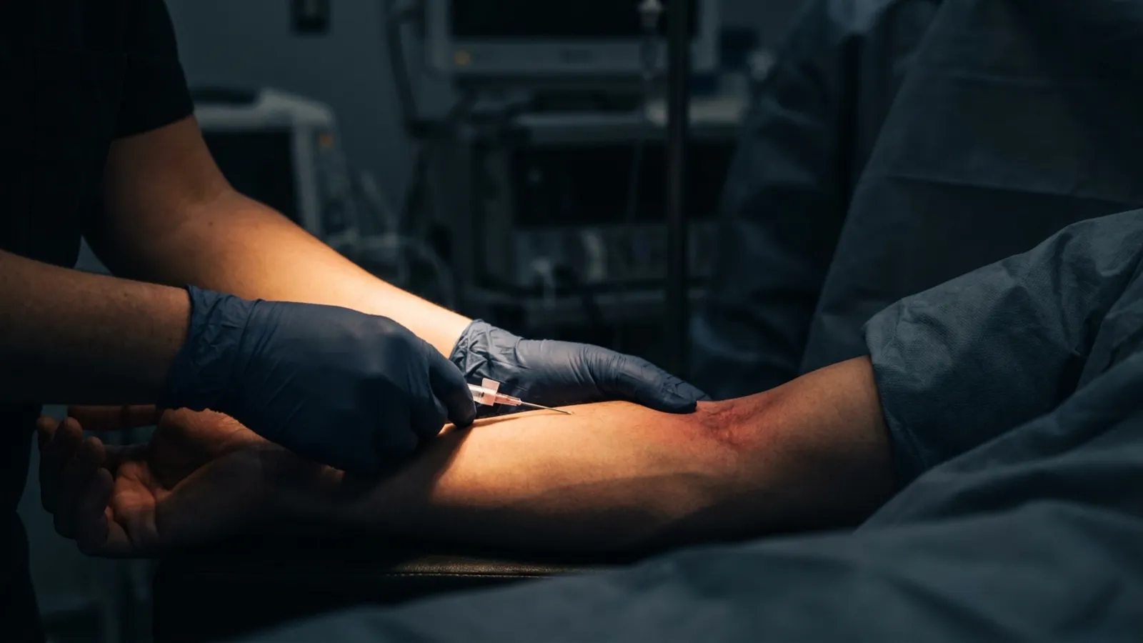
IV cannulation emergency department nurses perform daily is a different skill set than scheduled bedside access on a quiet med-surg floor. The patient is crashing, the room is loud, the chart is empty, and the line has to go in now. Master this and you become the provider others escalate to. Hesitate and the team works around you.
This is the field guide for ED vascular access. It covers site selection under time pressure, the two-attempt rule and the escalation pathway behind it, the role of ultrasound and external jugular access, the trauma activation playbook, and the psychology of working calmly when nothing around you is calm.
Why IV cannulation emergency department clinicians do is its own skill set
Emergency department vascular access happens under conditions that floor nursing rarely sees. Patients arrive dehydrated, hypotensive, intoxicated, agitated, or post-arrest. The history is a single sentence from EMS. The order is "two large-bore now." There is no time to coax a vein, warm an extremity, or wait for the next attempt to look better.
According to a 2024 systematic review and meta-analysis published in the International Emergency Nursing journal, ultrasound-guided peripheral IV cannulation by emergency nurses increases first-attempt success in difficult-access patients and reduces time to obtain access. The clinical implication is direct: ED nurses who add ultrasound to their toolkit measurably outperform those who rely on landmark technique alone.
Three things define ED cannulation as a distinct skill:
- Time pressure. A trauma activation patient needs IV access in the first 5 minutes. There is no luxury of a second careful attempt.
- Patient instability. Hypotension collapses peripheral veins. Agitation removes a still target. Hypothermia causes profound vasoconstriction. The vein you would normally hit is hiding.
- Limited information. No chart, no allergies, no history of previous access. You work with what you can see, palpate, or scan.
The ED reality: cannulation under chaos
A typical resuscitation room has 6 people moving around the patient, two monitors alarming, the airway team intubating, and a paramedic giving report at the same time you are starting a line. The cognitive load is enormous and your sympathetic nervous system is already engaged. This is where most missed sticks happen during IV cannulation emergency department staff perform under pressure, and the cause is rarely technique.
For broader context on managing your own state during high-pressure procedures, see our psychology of IV insertion guide. The principles are universal; the ED amplifies every one of them.
The clinical workflow that works under chaos:
- Look before you set up. Visual scan of both upper extremities while the team is still positioning the patient. You will identify your target faster than someone reaching for a tourniquet.
- Pre-stage your supplies. Two catheters open, flush ready, tegaderm peeled. The 30 seconds you save matter.
- Pick the right gauge for the indication. 16-18 gauge for trauma activation and resuscitation. 18-20 gauge for routine ED workup. Smaller for known difficult access. For a deeper read, see our IV catheter size selection guide.
- Stop talking. Once the catheter is in your hand, your mouth closes. Verbal multitasking competes with the motor planning your stick requires.
Site selection in the emergency department
Site selection for IV cannulation emergency department teams perform under time pressure follows a different hierarchy than scheduled access. Speed and reliability beat vein preservation in the immediate resuscitation, then preservation principles re-engage as the patient stabilizes.
Initial resuscitation hierarchy (first 10 minutes):
- Antecubital fossa. The basilic, cephalic, and median cubital veins are the first targets in any unstable patient. They are large, predictable, and tolerate large-bore catheters and rapid infusion.
- Forearm cephalic and basilic. Reliable backup if antecubital is occupied or unsuitable. Slightly slower but still appropriate for resuscitation.
- External jugular (EJ). Per the Journal of Emergency Nursing's published technique guidance, the EJ is an underutilized rapid-access site that does not require ultrasound when neck anatomy is favorable.
- Intraosseous (IO). When peripheral access fails or time does not permit further attempts, IO becomes the next escalation, not central line placement.
Stabilized patient hierarchy (post-resuscitation, awaiting admission):
- Distal forearm and dorsal hand. Once the patient is stable and the workup IV is the priority, return to standard preservation principles.
- Smaller gauge as appropriate. 20-22 gauge for routine fluids and labs.
- Avoid the antecubital for non-urgent access. Save the AC for IV contrast, blood draws, and the next emergency.
For the broader decision flowchart on when to reach for ultrasound, see our decision guide on when to use ultrasound for IV access.
The two-attempt rule and the ED escalation pathway
The two-attempt rule is more than a courtesy in the emergency department. It is the trigger for clinical escalation. Per the Society of Hospital Medicine position statement on ultrasound vascular access, patients with two failed landmark attempts are formally classified as having difficult intravenous access (DIVA) and warrant alternative techniques.
The ED escalation pathway:
- Attempt 1 (landmark technique). Visible or palpable vein, standard catheter, distal-to-proximal preference if not in resuscitation.
- Attempt 2 (landmark, different site). Different extremity or different segment. If you missed the right hand, look at the left forearm.
- Stop. Escalate. Two missed sticks is the trigger. Do not push for a third without changing your approach.
- Ultrasound-guided PIV (UGPIV). Studies show 84% success in patients with two prior failed landmark attempts when UGPIV is used.
- External jugular cannulation. When ultrasound is not available or when the EJ is the obvious target.
- Intraosseous (IO) access. When time is critical and peripheral options have failed. The Emergency Nurses Association's position statement on intraosseous access supports IO as the first alternative when peripheral access fails.
- Central venous catheter. Physician-placed, last resort for stable patients. Bypassed by IO in true emergencies.
For broader troubleshooting on why peripheral lines fail, see our guide on blown veins, causes, and prevention.
Comparison: standard PIV vs UGPIV vs EJ vs IO in the ED
| Technique | Time to Place | Success Rate (DIVA) | Best Use | Required Skills |
|---|---|---|---|---|
| Standard PIV | 30-90 seconds | 30-50% in DIVA | Visible/palpable veins, stable patient | Standard ED nursing |
| UGPIV | 2-5 minutes | 84% in DIVA after failed landmark | Difficult access, deep veins, planned access | Ultrasound certification |
| External jugular | 1-2 minutes | High when neck anatomy favorable | Rapid resuscitation, no ultrasound, hypotensive patient | EJ insertion training |
| Intraosseous | 30-60 seconds | Greater than 95% on first attempt | Critical resuscitation, failed peripheral, time-zero access | IO device training |
| Central line | 10-30 minutes | Procedure-dependent | Long-term ED access, vasoactive infusions, failed all alternatives | Physician/APP procedure |
Bottom line: In the unstable ED patient, the escalation order is two landmark attempts, then UGPIV or EJ, then IO. Central line is not your second move. It is the slowest option and bypassed by IO in any time-critical scenario.
Trauma activation: vascular access in the first 5 minutes
Trauma team activation is a defined workflow with vascular access at its center. According to the trauma team roles documented at McGovern Medical School, the IV nurse is positioned at the patient's right or left side with supplies pre-staged before the patient arrives. The expectation is two large-bore lines within the first 5 minutes of arrival.
The trauma IV protocol:
- Pre-arrival prep. Two 16-18 gauge catheters open. Pressure tubing connected to crystalloid. Tourniquet on the side you will work first.
- Visual scan during transfer. As the patient moves from the EMS gurney to the trauma bed, scan both arms. Pick your two targets before the team finishes positioning.
- First line: antecubital, large-bore. No exceptions for routine trauma activation. Large-bore AC IV first, every time.
- Second line: opposite extremity. Different side, same large-bore principle.
- Verbalize success or failure. "Right AC, 16, in." Or "Right AC missed, going left." The team needs to know.
- If both attempts fail. Call for IO. Do not attempt a third peripheral in an unstable trauma patient.
For the parallel skill set in flight medicine, see our guide on flight nurse IV requirements. The principles overlap; the venue makes everything harder.
Special ED populations
Different patient populations bend the standard ED approach in specific ways:
Pediatric ED: Pediatric trauma and resuscitation moves IO escalation forward in the pathway. After one failed peripheral attempt in an unstable child, IO is appropriate. Smaller gauges (22-24g) for stable pediatric workup. See our pediatric IV access tips guide for site selection and DIVA scoring.
Agitated and intoxicated patients: Restraint is a clinical decision the team makes together. Once the patient is appropriately restrained or sedated, vascular access becomes feasible. Forced cannulation on a moving target is unsafe for the patient and the staff. Wait for the right window.
Hypotensive and hypovolemic patients: Veins collapse under tourniquet release. Lower your insertion angle, advance slowly, and accept that the flash will be slower than expected. For the broader technique modifications, see our dehydrated patient IV access guide.
Post-cardiac arrest patients: Vasoconstriction and clinical instability make peripheral access difficult. EJ and IO are often the right initial moves. Standard peripheral attempts can wait until ROSC and stabilization.
Patients with prior IV drug use: Vein scarring at the standard antecubital and forearm sites limits options. Look at uncommon sites (dorsal hand, lower forearm radial) and consider UGPIV early.
The psychology of ED cannulation
The technical skill is half of ED vascular access. The other half is what your central nervous system is doing while you place the line. A heart rate above 120, narrowed visual field, and shaking hands are not personal failings. They are predictable physiologic responses to high-stakes performance under observation.
The providers who succeed in ED cannulation have trained their nervous system to stay regulated when the room around them is not. We teach this connection at VeinCraft Academy as part of our psychology-first curriculum. The mental game is the technique, not separate from it.
The identity to cultivate is the nurse the trauma team trusts to get the line. The one who walks into the room, scans the arm, picks the target, and places the catheter without performance commentary. That confidence is built, not born. It comes from training, repetition, and a framework for handling complexity.
For the broader career trajectory ED IV competence opens, see our guide on nurse career advancement through IV skills. ED competence is the gateway to flight, ICU, and critical care roles.
How VeinCraft trains for emergency department vascular access
Level 2: The Craft covers the clinical decision-making behind ED-level access: hard-stick technique, ultrasound-guided peripheral access, EJ cannulation, and the psychology of working calmly under pressure. Students practice under the observation of credentialed clinical instructors with active field experience. Mastery-based progression means you advance when you demonstrate competence under simulated stress, not when the clock runs out.
If you are new to IV cannulation, start with Level 1: The Method at $199. The 8-hour intensive builds the foundation that makes Level 2 productive. The bundle (Master the Craft) at $449 saves $49 and includes a free practice kit. Enroll in the next cohort when you are ready to be the nurse the trauma team calls for.
Frequently asked questions
What gauge IV is used for trauma activation in the emergency department?
A 16 or 18 gauge catheter is standard for trauma activation, placed in the antecubital fossa for rapid large-volume infusion. Two large-bore lines are the protocol expectation in the first 5 minutes of arrival. Smaller gauges (20-22) are reserved for stable ED workup once resuscitation needs are addressed.
What is the two-attempt rule in ED IV access?
The two-attempt rule is a clinical standard that two failed landmark IV attempts triggers escalation to alternative techniques rather than continued peripheral attempts. Patients meeting this criterion are formally classified as having difficult intravenous access (DIVA). The next steps are ultrasound-guided peripheral IV, external jugular cannulation, or intraosseous access depending on patient stability and equipment availability.
When should an ED nurse use intraosseous instead of peripheral IV?
Intraosseous access is appropriate when peripheral attempts have failed, when time-critical resuscitation does not permit additional peripheral attempts, or when patient anatomy makes peripheral access unrealistic. Per Emergency Nurses Association guidance, IO should be considered as the first alternative when peripheral access cannot be rapidly obtained. IO placement is faster than central line placement and bypasses the need for physician procedure in most settings.
Can ED nurses place external jugular IVs?
Yes, in most jurisdictions and institutions. External jugular IV cannulation is within RN scope of practice in emergency departments with appropriate institutional training. The EJ is a peripheral vein and the technique is similar to standard PIV with attention to neck positioning and patient airway management. EJ access is a valuable skill when ultrasound is unavailable and rapid access is required.
How does ED IV cannulation differ from floor nursing?
IV cannulation emergency department teams perform operates under time pressure, patient instability, and limited information that floor nursing rarely encounters. ED nurses must master a broader access toolkit including ultrasound, EJ, and IO escalation. Site selection prioritizes speed and reliability during resuscitation, then shifts to vein preservation as the patient stabilizes. The cognitive load is significantly higher and the psychology of cannulation under chaos is its own skill.
Should ED nurses learn ultrasound-guided IV placement?
Yes. Ultrasound-guided peripheral IV (UGPIV) is rapidly becoming a baseline expectation for IV cannulation emergency department staff perform daily. Studies show UGPIV by trained ED nurses achieves 84% success in patients with two prior failed landmark attempts, reduces time to access, and reduces the need for physician intervention. Most academic and community EDs now offer or require UGPIV training for nursing staff.
VeinCraft Academy is a mastery-focused IV cannulation training program for healthcare professionals. All instruction is delivered by credentialed clinicians with active field experience. VeinCraft Academy is a RevivaGo Company.